Meghan Root's E-Portfolio
Research Pathway
From Technique to Treatment
Cell and Molecular biology lab (Biology 302 lab) was one of my favorite classes I have taken thus far at USC. In biology 302 lab we spent the majority of the semester looking at the HL-60 cell line. This cell line is unique because it is a myeloblastic cell line, meaning the cells can differentiate into other cells. This is important because with the right growth factors the cells can differentiate an unlimited number of times and into many different types of cells.
We used this cell line and looked at many different aspects of the cells, specifically we looked at differentiation, gene expression, and protein content. The lab where we looked at proteins within the cells was the most interesting to me. We used Western Blots and SDS-PAGE analysis to look at the different proteins that were being expressed. We then used the information we gathered to make conclusions about the cell line. PMA and DMSO are both different growth factors that bind to receptors within the cell and lead to changes in gene expression. MMP-9 is a class of enzymes that are involved in the degradation of the extracellular matrix. For example, in my I hypothesized that the cells treated with PMA will have higher protein content and contain more active MMP-9 than in the cells treated with DMSO. In order to figure out the specific protein content of the cells we used SDS-PAGE and Western Blot analysis. I thought these experiments were so interesting because we were able to take a sample of a cell culture, place it on gel and separate the proteins by electrophoresis. We would then end up with picture of what the protein content looks like, and be able to learn about what is going on within the cells.
Due to the information and research techniques I learned in Biology 302 lab, I was inspired to seek out more research opportunities. I was a research assistant at Johns Hopkins in the Eberhart, Rodriguez and Raabe Pathology Laboratories. During my time in the lab I helped fellow Jeffrey Rubens M.D. with his research on atypical rhabdoid tumors (ATRT tumors) of the brain. ATRT tumors are cancerous, fast-growing rare tumors of the brain and spinal cord. ATRT tumors often result from abnormalities in a gene that makes proteins to stop tumor growth. In this tumor the gene doesn’t function properly therefore, the tumor growth is uncontrolled. Fellow Jeffrey Rubens M.D. was using Western Blots to look at the proteins within the ATRT tumor cells after they had been treated with Cisplatin, which is a chemotherapy medication or DON, which is a potential therapeutic. Having the prior knowledge of Western Blots from 302 lab enabled me to better utilize my time over the summer. I was more confident in my ability to perform a Western Blot and therefore, I was able to focus more of my time on learning about the specific potential therapies for the ATRT tumors. Fellow Jeffrey Rubens M.D. is continuing this research and working towards published within the coming months.
As an aspiring medical professional, my time at Johns Hopkins over the summer has opened my eyes to the sheer importance of research. The idea that learning something as simple as the technique of a Western Blot allows you to look at the protein content of the cells from a very rare and deadly pediatric brain tumor and potentially come closer to figuring out a way to treat it, really blows my mind. Prior to my research experience I was aware of the necessity of research, just as many people are, but I don’t think it fully hit me until I was able to experience it for myself and actually see the patients we were working to help. I think this experience will push me to work harder in my medical career and strive to help as many people as I possibly can.

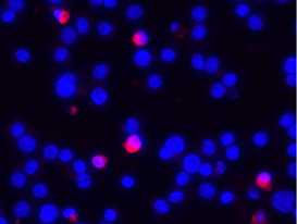
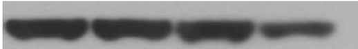
The images to the left are examples of a Brdu assay. This assay is used to look at cell proliferation, or cell growth which is normally exhibited at a higher rate in tumors. In the lab we used this assay to determine the growth within the ATRT tumors following treatment with certain drugs. The cells that are highlighted bright blue are the ones that are living and the cells that are faded, or barely visible are the cells that are dead. The picture above is actin in a Western Blot. This is used to create the control to test the experimental cells against. I was able to explore assays such as the Brdu assay due to the confidence I had in lab technique die to Biology 302 lab.